���̉p���́u���ۃ��[�N�V���b�v�@�A�W�A�ɂ����邶��x��v
�i�����w��w�� ���ێЉ��w�u�����ی��w�̈� ����������Á@05,12�j��
�������̂ł��B �i���z�����a�@�@���쎡�a�j |
| I am a general physician. I have an interest in occupational and environmental health. I present the medical support for pneumonoconiotic patients in the local community. I feel very honored to give you today's workshop. Professor Kusaka, I express gratitude from my heart. I am making a speech with 4 points. First, the chest x-ray diagnosis of pneumonoconiosis. �@The comparison with our hospital diagnosis and the diagnosis of the Fukui Labor Office Second, The transition of the respiratory function during observation periods, an average of seven years. Third, The complications of pneumonoconiosis and the result of the bacterial culture of sputum. The respiratory failure accompanied by pneumonoconiosis advances gradually.�@ Therefore, Home oxygen therapy is needed. Finally My patient's prognosis. According to the official announcement by the Ministry of Health, Labor and Welfare, the total number of patients with pneumonoconiosis was 9160 in 2002. Among then, 104 patients live in Fukui prefecture. More than half of them were treated at my hospital. The number of patients studied is 109�i1995 �` 2005). They are retired now. 83 patients are alive. 26 patients are dead.�@The mean age is 71.7. 106 patients have silicosis and 3patients have coal mine workers. This slide is the work types. The exposure period was an average of 25.9 years. Most of them were tunnel workers.�@ Subsequently there were also a numbe of zinc mine, stone masonry, and coalmine workers.
It is important for pneumonoconiotic patients to stop smoking..90 percent of my patients had a habit of smoking before. �@I recommend my patients a non smoking program. �@According to my program, 80% of them have stopped smoking. �@However I am suspicions about whether this number is reliable or not, because it is well known that a patient may not talk honestly in front of a doctor. In Japan there are a large number of tunnels. �@In Fukui Prefecture there is the Hokuriku tunnel, which used to be the longest in Japan. �@ I have many patients who worked there. I also have many patients who were worked for the zinc mining company. It was NAKATATU mine in Izumi Village. This mine had a large output of zinc, and had the second most products in Japan. They finished mining in NAKATATU 23 years ago. I was born and lived there until I went to the junior high school close to NAKATATU in Izumi Village. 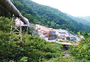 �@ �@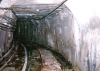 (Nakatatu�@Mine) This is the picture of tunnel workers who were drilling a rock 30 years ago .�@The workers didn't put on the antidust mask except for one person. �@Pneumonoconiosis of tunnel workers is more prevalent than that of zinc mine workers. 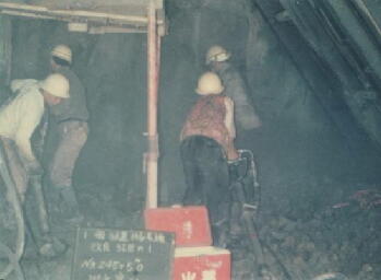 In Japan, according to the pneumonoconiosis law, the workers exposed to mineral dust must have a medical checkup once a year. Doctors indicate the diagnosis and degrees of pneumonoconiosis on documents, and present them to the labor office. �@The specialist team of the Fukui labor office discusses the documents. Finally the Classification for supervision of pneumoconiosis is determined. In the pneumoconiosis law, radiographic appearances of pneumoconiosis shall be classified into the four categories of Categories I to IV as shown in the center column the table of this slide. ILO classification is shown in the right column the table of this slide. Categories 1 to 3 are those which are deemed containing small opacities and containing no large opacities Categories 4 are those which are deemed containing only large opacities.
This slide shows the difference in a chest x-ray diagnosis between our hospital and the Fukui labor office. �@We are in agreement in about 80 percent of the case (Deep red column). �@We are not in agreement 20%. Blue column express overestimation in18%. Pink. column express underestimation in 4%. Did we overestimate or did the labor office underestimate? I will explain the difference.
This slide illustrates for every Japanese Category in the chest x-ray. I compared the category which was determined by the Fukui labor office with the category by my hospital. This is our category and this is the category by the Fukui labor office. There is a big difference in category 4. This difference is caused by large opacities I detected large opacities, while the Fukui labor office did not detect large opacities.
Large opacities are divided into Categories A, B, and C Category A is one or more opacities whose combined diameters do not exceed 5 cm Category B is one or more opacities, larger and more numerous than A, whose combined area does not exceed the equivalent of the right upper zone Category C is one or more opacities whose combined area is larger than the equivalent of the right upper zone I'll show you two cases. The first case is a male tunnel worker who is 71. I diagnosed the small opacities as 2 and large opacities as A. The labor office diagnosed the small opacities as 1, and they detected no large opacities. In this case, I think it is difficult to detect large opacities  This is the same patient's chest CT. The CT clearly detected the large opacities both in a lung field level and a mediastinum level   Second case is a male tunnel worker who is 70. I diagnosed the small opacities as 2 and the large opacities as A. The labor office diagnosed the small opacities as 2, and detected no large opacities. I think it was easy to detect the large opacities, although they had overlapped with the collarbone. What do you think of this?  This is the same patient's chest CT.�@ Large opacities are detected clearly. In the Labor Office they take the chest X-ray only, to diagnosis pneumonoconiosis. Usually we take both chest X-ray and CT for all the patients, so we can detect large opacities easily. This would be the reason for the difference.   Next, this is the transition of the respiratory function. % vital capacity was 89.5% seven years ago and 79.8% now. �@Vital capacity is decreasing gradually.
On the other hand, the %of FEV1.0 is not decreasing. In our study, many patients have the mixed type pulmonary dysfunction. The % of vital capacity shows the better marker of pulmonary dysfunction rather than that of the % of FEV1.0.
Arterial Blood Partial Oxygen Pressure was 80.9torr six years ago and 73.9torr now. Statistically, this is a significant difference. Hypoxemia is advancing gradually.
This is the graph of the complications of pneumoconiosis. 90 percent of the patients have secondary bronchitis. We have also found of pneumothorax and tuberculosis in our patients. Secondary bronchitis is terminology persistently used in the pneumoconiosis law. The Japanese pneumoconiosis law defined this as follows. When we collect dirty sputum from bacterial infections within one hour after a patients waking early morning, we find 3ml more over, this should be recognized as a complication from pneumoconiosis
I examined sputum of 26 bronchitis patients every month for one year. The total number of samples is 253. �@A bacterial culture of sputum was performed. Pathogenic bacteria were detected in 47% of the samples. I considered bacterial bacillus classification. �@Most of them were Klebsiella pneumoniae and Pseudomonas aeruginosa.�@In decreasing order the other bacterial bacillus, Hemophilus influenzae, Pneumococcus , serratia marcescens, MRSA. Recently, atypical mycobacterial infection has been detected
Silicosis is an irreversible condition, with no cure. Treatment options currently focus on alleviating�@the symptoms and preventing complications. These include is Stopping tobacco smoking. Cough suppressants. Antibiotics and antitubercular agents. Chest physical therapy to help the bronchial drainage of mucus. Oxygen administration to avoid hypoxemia. Bronchodilators to facilitate breathing. �@We have many types of Bronchodilators for inhalation. I prescribe these bronchodilators for my patients. My patients are taking medication, chest physical therapy and a non-smoking program. However, respiratory failure is advancing gradually. The number of patients who need home oxygen therapy is increasing. I have seven patients who are taking home oxygen therapy now. I think the risk factors are emphysema, bronchitis, pneumonia, pneumothorax, and lung cancer. He is a male tunnel worker who is 74. He suffered from pneumothorax 4 times. Pao2 shows 53.5 torr in room air, %VC shows 57% now. He is taking home oxygen therapy for a whole day. �@Whenever he takes a bath, he feels severe shortness of breath. �@The spo2 falls into below 80% immediately.He sued several company because he is got pneumoconiosis . He said like this; I worked in tunnels everywhere in Japan. �@I had to retire when I was 50 years old, because of the shortness of breath.�@�@It was not use for him to put on the antidust mask, because the mask got dirty with dust in a few miniatures.�@The company neglected workers' medical checkup. �@The management supervisor of the labor office did not investigate by enter the tunnel. I cant live so long. �@I didn't want to anyone to get pneumoconiosis any more. 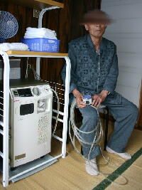 �@ �@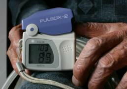 I analyzed the cause of death of 26 patients' examples. Most were from respiratory failures and, subsequently lung cancer. Others include malignant tumors, and tuberculosis.
CONCLUSION Comparison with the chest x-ray diagnosis of our hospital and the diagnosis of the Fukui labor office. The rate of coincidence was about 80 percent. Although large opacities is easy to be detected in chest CT, to overlook in chest x-ray. Although %Vital capacity and Arterial Blood Partial Oxygen Pressure is fallen in average observation period, it didn't fall %FEV1.0. 93% of patients were suffered from bronchitis.I detected bacteria from the 47% of the samples. Klebsiella pneumoniae and Pseudomonas aeruginosa were the most detected. The patients who need home oxygen therapy are increasing. The most common cause of death is respiratory failures. Thank you for your attention. |